This months focus will be on ECG’s with some STEMI equivalents and revision on other Infarct patterns. We’ve had some great ECG’s amongst Ryde crews recently so this should be a good refresher with consolidation of some ECG patterns to keep an eye out for. If you’re still with me by the end have a go of the two ECG tests and check your knowledge. They are quite hard but there’s some great learning points to be had by trying them. After the ECG tests there are also a couple of skills: if you get some time on station go to the training room and attempt the skill stations set up there.
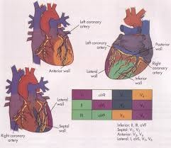
To kick off we’re going to do a quick review of coronary artery anatomy:

Coronary artery anatomy is individual, but the images here demonstrate a pretty accurate representation of the layout in the majority of people. Take note of the arteries that feed the leads of our 12 lead, as this will become relevant with the following ECG patterns.
STEMI Equivalents
The following are “STEMI equivalents”, the Dewinter pattern, Wellen syndrome and aVR elevation with widespread horizontal depression. Recognising one of these in a patient and subtly changing your treatment or activating the cath lab may save their lives. The following quote is from the British Cardiovascular Society:
“It is not widely recognised that the potentially devastating forms of STEMI (STEMI-equivalent) may present electrocardiographically quite subtly without classical ST elevation. These changes as part of an appropriate presenting syndrome form no part of the classic referral into a PPCI pathway. Yet prompt recognition of them by every physician dealing with acute admissions should trigger referral for urgent coronary angiography with an appropriately timely reperfusion strategy.”
DeWinters T Waves:The pattern in the image above of a deep ST segment depression in leads V2-4 is a highly sensitive one for a proximal LAD(left anterior descending) artery occlusion.
Watch this video for a case presentation and description of this indicator of a occlusion requiring cath lab activation:
http://ekgumem.tumblr.com/post/19233090476/what-are-de-winter-t-waves-episode
Wellens Syndrome:
Have a read of the following on the importance of recognising inverted/biphasic T waves in V2-V4, note in particular the rates of AMI in the days following Wellens syndrome being seen on ECG.
http://www.academiclifeinem.com/wellens-syndrome-is-it-on-your-radar/
aVR ST segment elevation and widespread ST segment depression:
There is a good example of this type of STEMI equivalent on the plant room whiteboard. The aVR elevation, especially when greater than the elevation in V1 and in the presence of inferolateral ST depression is highly specific for Left Main Coronary Artery(LMCA) occlusion. The mortality associated with this type of occlusion without treatment is huge, 70-85%, so this is one to be vigilant for.
A case presentation:
http://lifeinthefastlane.com/another-widow-maker/
And some more examples and information on this pattern:
http://lifeinthefastlane.com/ecg-library/lmca/
Sgarbossa:
A good rundown on how to differentiate the presence of a critically occluded coronary artery in the presence of a LBBB or paced rhythm:
Right Ventricular Infarct:
RVI is present in up to 40% of Inferior MI’s, but why should this matter to us if we’ve already spotted the Inferior ST segment elevation? Check out this great video for the answer!
http://ekgumem.tumblr.com/post/17948681579/how-to-diagnose-right-ventricular-mi-episode
Doing lead V4R to check for RVI, particularly in the setting of an Inferior MI, is simple and quick to do. Take V1 and move it to the same position as V4 but on the Right side of the chest and then perform another 12 lead. Don’t forget to label the changed lead on the ECG printout to save any confusion.
Posterior MI:
Lastly a quick note on posterior MI’s. Mostly associated with inferior infarctions(Remember the RCA feeds across the Right side, inferior wall and the posterior wall) Isolated Posterior MI’s are very rare, constituting less than 3% of all AMI. The key to this is that ST depression in V1-V3 mirrors the posterior wall, so if you flip the ECG upside down it will show the ST elevation. To confirm the presence of posterior wall infarction pull leads V4-V6 off and place them as follows in V7-V9. The ST depression you spotted in the septal leads will be mirrored by ST depression in these leads if there is indeed a posterior infarct.
Still with us? Great, here’s two ECG tests that will fry your brain and are great to consolidate some of what we’ve covered this month:
The first one will give you a transcript of your accuracy at the end and compare you with ED docs, all docs, cardiologists and the computer algorithim. It’s based on a study by McCabe et al where these same 36 ECG’s were interpreted by clinicians from all these backgrounds.
https://script.google.com/macros/s/AKfycbyZEqQ5jxNsDl7Nfijw77PIEdOjY4BSbcx4mER55Ffqh4NjamE/exec
Next up is the University of Maryland Registrar’s ECG exam, try it then check your answers on the embedded video:
1405071322_2014_UMEM_ECG_Competition
MARYLAND ECG TEST
Skills:
In the training room you will find 4 skill stations:
1- Patient trapped by compresion, draw up Sodium Bic and Calcium Gluconate
2- Paediatric asthma, draw up IM adrenaline dose
3- Paediatric naloxone dose
4- 15 lead ECG dot location(posterior and RV)
Get in and have a go! Don’t forget to log time spent to gain CTP points.


